
| REF | DESCRIPTION | SAMPLES PER GEL | TEST PER KIT |
|
|
|---|---|---|---|---|---|
| SRE645K | CSF ISOELECTROFOCUSING KIT | 2 | 20 | ||
| SRE622K | 6 | 60 |
The Interlab CSF Isoelectrofocusing Kit SRE622K is intended for identifying oligoclonal banding in paired serum and CSF using isoelectric focusing and immunoblotting.
This technique is considered “The Gold Standard” method for the determination of intrathecal IgG synthesis in the clinical diagnosis of multiple sclerosis. In fact, isoelectric focusing is the most sensitive method for the detection of oligoclonal bands is serum and CSF (0.040mg/dl).
The procedure includes Isoelectrofocusing on agarose gel using the Interlab instruments and manual immunoblotting steps. Isoelectrofocusing on agarose gel has the purpose to fractionate the proteins in the CSF and serum samples. The immunoblotting steps have the purpose to transfer proteins on the transfer membrane. This transfer membrane is processed to detect IgG oligoclonal bands and to demonstrate the difference, or lack of, in the distribution of IgG in the CSF and serum of the same patient. The immunofixation with labeled anti-IgG antiserum permits to detect only the true IgG oligoclonal banding at increased sensitivity of detection so that the analysis can be generally performed on unconcentrated CSF.
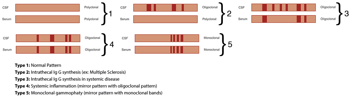
The IgG immunofixation patterns of CSF and serum from the same patient are then visually compared. This allows detection of oligoclonal banding that represents intrathecal synthesis of immunoglobulins. Five different patterns may be seen after the isoelectric focusing (see Fig. below):
Type 1: Normal CSF, no band present in the CSF.
Type 2: Intrathecal IgG synthesis. CSF with restricted oligoclonal bands not seen in the serum, found in multiple sclerosis.
Type 3: Intrathecal IgG synthesis: CSF with restricted oligoclonal bands with additional bands seen in both the CSF and serum. It is found in multiple sclerosis and brain inflammation in systemic disease, for example, sarcoidosis.
Type 4: Identical oligoclonal bands in the CSF and serum. Monoclonal bands found in systemic inflammation, for example, Guillain-Barrè syndrome.
Type 5: Monoclonal bands in both the CSF and serum. It is found in myeloma or monoclonal gammopathy of uncertain significance
Reagent Preparation:
For the preparation of the reagents refer to the procedure.
Sample Preparation:
Neat CSF samples. The concentration of IgG in the paired serum samples should be adjusted to the same level of the CSF samples using distilled water.
Sample Storage & Stability:
Serum/CSF: Fresh serum and CSF samples. If needed: 1 week at 2 to 8°C, 1 month at -20°C