


| REF | DESCRIPTION | SAMPLES PER GEL | TEST PER KIT |
|
|
|---|---|---|---|---|---|
| SRE651K | SERUM AND CONCENTRATED URINE/CSF IMMUNOFIXATION ACID VIOLET STAIN | 1 | 10 | ||
| SRE627K | 2 | 20 | |||
| SRE628K | 4 | 40 | |||
| SRE639K | 6 | 60 |
The new Immunofixation Electrophoresis (IFE) kits are intended to be used for qualitative immunological identification of monoclonal components in human serum and in concentrated urines/CSF. The use of high sensitivity staining solution (Acid Violet) and the new enhanced formulation guarantees speed and high sensitivity. Thanks to the new Easy Interlab G26 & Pretty Interlab the Immunofixation procedure is extremely fast and user friendly and in just 43 minutes the first gel results is completed. The kits have been designed for use with the fully automated instruments Easy Interlab G26 & Pretty Interlab.
Reagent Preparation:
All reagents ready to use except the washing solution to be diluted 50ml to a final volume of 1L with distilled water.
Sample Preparation:
Neat serum samples. Concentrated urines to a final total protein value of about 5 g/L.
Sample Storage & Stability: Serum:
۱ week at 2 to 8 °C Urine/CSF: 1 week at 2 to 8 °C, and 1 month at – ۲۰°C
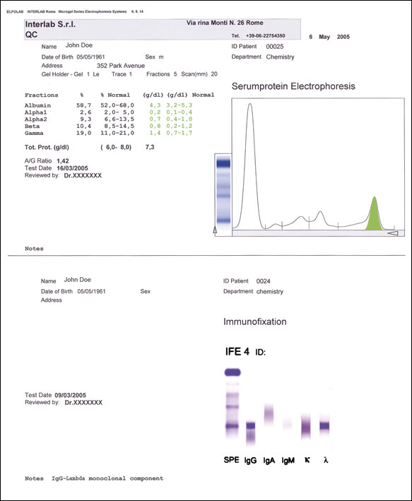
The neoplastic proliferation of single clones of plasma cells, a condition also termed monoclonal gammopathy, causes the abnormal synthesis of monoclonal immunoglobulins, namely a biochemically homogeneous group of immunoglobulins consisting of one single type of heavy chain and light chain. These monoclonal immunoglobulins are also called paraproteins and are frequently associated with a broad heterogeneous group of plasma cell dyscrasias.
In most cases these paraproteins produce one or more sharp bands in the electrophoretic patterns obtained from serum and/or urine samples. Although monoclonal components are typical of myeloma, they also appear on the electrophoretic pattern of patients suffering from other conditions like infections and auto-immune diseases. Occasionally their presence is observed in a few benign conditions in elderly individuals.
These bands are generally described as ‘suspected monoclonal components’, and their biochemical identity needs to be further investigated with sensitive and specific electrophoretic methods. Confirmation of the presence of a monoclonal immunoglobulin, together with the characterization of the immunoglobulin type (for example IgG, k or IgM, λ) are of fundamental importance for the definitive diagnosis. Immunofixation electrophoresis (IFE) is a laboratory method used to define the biochemical identity and homogeneity of immunoglobulins, when suspected monoclonal components are detected in protein electrophoretic patterns of biological fluids. IFE Acid Violet stain combines the resolution of protein fractions by electrophoresis with the specific recognition of molecules using antibodies raised against heavy chains of human immunoglobulins (IgG, IgM, and IgA), and their light chains, kappa and lambda. The binding between the specific antibody and the monoclonal immunoglobulin results in the formation of a band of precipitate in the corresponding lane that identifies the type of immunoglobulin, either heavy chain and/or light chain. The use of acid violet, a very sensitive stain for proteins, provides improved quality of the electrophoretic results, for a better visual inspection of the patterns.